Signaling Systems in Development
|
| Conserved Signaling Systems
Used in the Development of Animals |
| SIGNALING PATHWAY |
LIGAND FAMIILY |
RECEPTOR FAMILY |
EXTRACELLULAR MODULATORS |
|
|
| Receptor tyrosine kinase (RTK) |
EGF |
EGF receptors |
Argos |
|
FGF (Branchless) |
FGF receptors (Breathless) |
|
|
ephrins |
Eph receptors |
|
| TGFb
superfamily |
TGFb |
TGFb
receptors |
chordin (Sog), noggin |
|
BMP (Dpp) |
BMP receptors |
|
|
Nodal |
|
|
| Wnt |
Wnt (Wingless) |
Frizzled |
Dickkopf, Cerberus |
| Hedgehog |
Hedgehog |
Patched, Smoothened |
|
Sonic HedgeHog
- FUNCTION:
binds to the patched (ptc) receptor, which functions in association with smoothened
(smo), to activate the transcription of target genes. in the absence of shh, ptc
represses the constitutive signaling activity of smo . also regulates another target, the gli
oncogene. Intercellular signal essential for a variety of patterning events during
development: signal produced by the notochord that induces ventral cell fate in the neural
tube and somites, and the polarizing signal for patterning of the
anterior-posterior axis of the developing limb bud. displays both floor plate-and
motor neuron-inducing activity.
- SUBCELLULAR
LOCATION: the c-terminal peptide diffuses from the cell, while the n-terminal
peptide remains associated with the cell surface. is also secreted in either cleaved or
uncleaved form to mediate signaling to other cells
Patched - SHH Receptor
- FUNCTION:
acts as a receptor for sonic hedgehog (shh), indian hedgehog (ihh) and desert
hedgehog (dhh). Associates with the smoothened protein (smo) to transduce the hedgehog's
proteins signal. Seems to have a tumor suppressor function, as inactivation of this
protein is probably a necessary, if not sufficient step for tumorigenesis.
- DEVELOPMENTAL STAGE: in the embryo, found in all major target tissues
of sonic hedgehog, such as the ventral neural tube, somites, and tissues surrounding the
zone of polarizing activity of the limb bud.
Smoothened (co-receptor)
- FUNCTION:
g protein-coupled receptor that probably associates with the patched protein
(ptch) to transduce the hedgehog's proteins signal. binding of sonic hedgehog (shh) to its
receptor patched is thought to prevent normal inhibition by patched of smoothened (smo).
- SUBCELLULAR
LOCATION: integral membrane protein. belongs to family fz/smo of g-protein
coupled receptors.
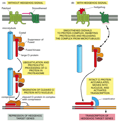
(A) In the absence of Hedgehog, the Patched receptor inhibits Smoothened probably
by promoting the degradation or intracellular sequestration of Smoothened. The Ci protein
is located in a protein complex and is cleaved to form a transcriptional repressor, which
accumulates in the nucleus to help keep Hedgehog target genes inactive. The protein
complex includes the serine/threonine kinase Fused, the anchoring protein Costal (which
binds the complex to microtubules), and the adaptor protein Suppressor of Fused. (B)
Hedgehog binding to Patched relieves the inhibition of Smoothened, which now signals to
the protein complex to stop processing Ci, to dissociate from microtubules, and to release
the unprocessed Ci so it can accumulate in the nucleus and activate the transcription of
Hedgehog-responsive genes. Most of the molecular events in the pathway are unknown.
Wnt proteins
- DEFINITION A family of
highly conserved secreted signaling molecules that regulate cell-to-cell interactions
during embryogenesis. Wnt (wingless) genes and Wnt signaling are also implicated in
cancer. Insights into the mechanisms of Wnt action have emerged from several systems:
genetics in Drosophila and Caenorhabditis elegans; biochemistry in cell
culture and ectopic gene expression in Xenopus embryos. Many Wnt genes in the mouse
have been mutated, leading to very specific developmental defects.
- RECEPTOR - FRIZZLED As currently
understood, Wnt proteins bind to receptors of the Frizzled family on the cell surface.
Through several cytoplasmic relay components, the signal is transduced to beta-catenin,
which then enters the nucleus and forms a complex with TCF to activate transcription of
Wnt target genes. . Wnt signaling has been discussed in and excellent web page maintained
by Roel Nusse
- CO-RECEPTOR protein is related to the low density
lipoprotein (LDL) receptor protein and is therefore called LDL-receptor-related protein
(LRP). It is uncertain how Frizzled and LRP activate Dishevelled, which relays the signal
onward.
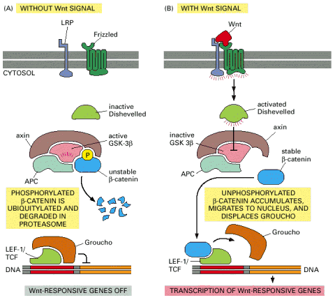
- ACTION (below) (A) In the absence of a Wnt signal, some b-catenin is bound to the cytosolic tail of cadherin proteins (not
shown) and any cytosolic b-catenin becomes bound by the
APC-axin-GSK-3b degradation complex. In this complex, b-catenin is phosphorylated by GSK-3b,
triggering its ubiquitylation and degradation in proteasomes.Wnt -responsive genes are
kept inactive by the Groucho corepressor protein bound to the gene regulatory protein
LEF-1/TCF. (B) Wnt binding to Frizzled and LRP activates Dishevelled by an unknown
mechanism. By an equally mysterious mechanism, which requires casein kinase 1 (not shown),
this leads to the inactivation of GSK-b3 in the degradation
complex. As a result, the phosphorylation and degradation of b-catenin
is inhibited, and b-catenin accumulates in the cytoplasm and
nucleus. In the nucleus, b-catenin binds to LEF-1/TCF,
displaces Groucho, and acts as a coactivator to stimulate the transcription of Wnt target
genes.
Wnt and Hedgehog signaling Compared
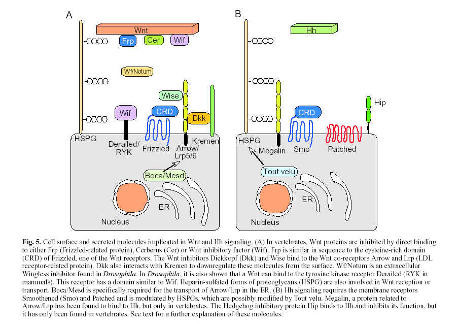
From Nusse Development
130, 5297-5305 2003 The Company of Biologists Ltd
Fibroblast Growth Factor
Fibroblast Growth Factor (FGF) was originally identified as acidic or basic, and
we now know there to be at least 17 different FGFs. These protein growth factors are bound
by 4 different cell membrane receptors (FGFR1-4). FGFRs belong to the tyrosine kinase
receptor family.
- FUNCTION:
the heparin-binding growth factors are angiogenic agents in vivo and are potent
mitogens for a variety of cell types in vitro. there are differences in the tissue
distribution and concentration of these 2 growth factors.
FGF Receptor
- FUNCTION:
receptor for basic fibroblast growth factor.
- CATALYTIC
ACTIVITY: ATP + protein tyrosine = ADP + protein tyrosine phosphate.
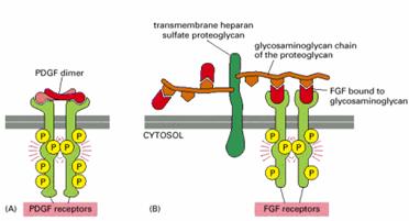
Platelet derived Growth Factor (PDGF) Receptor Compared with FGF receptor
TGF-beta
Transforming Growth Factor-beta (TGF-b) is a
protein growth factor acting through membrane receptors to signal proliferation and
differentiation in many different cell types. There are a large number of different
proteins belonging to this family. They can also interact with other growth factors to
stimulate or inhibit their action. Bone morphogenic protein (BMP) functions
to induce cartilage and bone formation and belongs to the Transforming Growth Facto -beta
(TGF-beta) family.
TGF-beta Receptor
TGF-beta receptors (Type I and Type II) dimerize to form an active threonine
serine kinase which autophosphorylates and recruits and activates Smads.
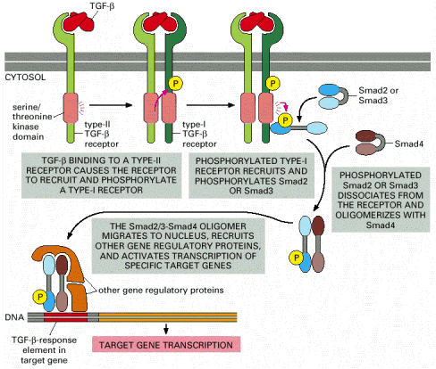
Note that TGF-b is a dimer and that Smads open up to
expose a dimerization surface when they are phosphorylated. Several features of the
pathway have been omitted for simplicity, including the following: (1) The type-I and
type-II receptor proteins are both thought to be dimers. (2) The type-I receptors are
normally associated with an inhibitory protein, which dissociates when the type-I receptor
is phosphorylated by a type-II receptor. (3) The individual Smads are thought to be
trimers. (4) An anchoring protein (called SARA, for Smad anchor for receptor
activation) helps to recruit Smad2 or Smad3 to the activated type I receptor by
binding to the receptor, to the Smad, and to inositol phopholipid molecules in the plasma
membrane. (5) The function of certain Smads is regulated by enzymes that enhance their
ubiquitylation and thereby their degradation.
Notch
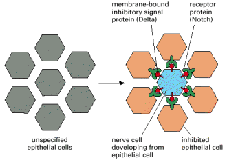
- FUNCTION lateral inhibition of adjacent cells influencing their
fate.When individual cells in an epithelium begin to develop a specific fate , they signal
to their neighbors not to do the same. This inhibitory, contact-dependent signaling is
mediated by the ligand Delta that appears on the surface of the differentated cell and
binds to Notch proteins on the neighboring cells. In many tissues, all the cells in a
cluster initially express Delta and Notch and a competition occurs, with one cell emerging
as winner, expressing Delta strongly and inhibiting its neighbors from doing likewise. In
other cases, additional factors interact with Delta or Notch to make some cells
susceptible to the lateral inhibition signal and others deaf to it.
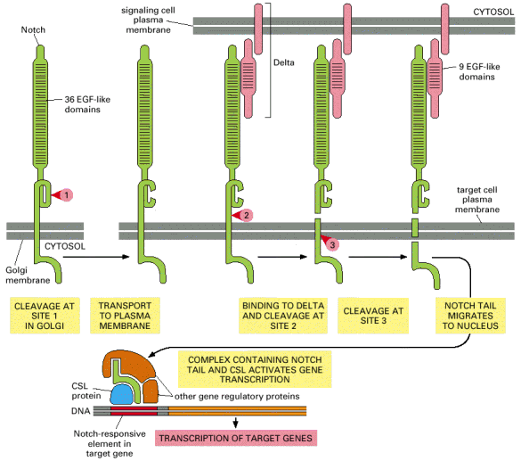
- ACTIVATION (Below) The numbered red arrowheads indicate the
sites of proteolytic cleavage. The first proteolytic processing step occurs within the trans
Golgi network to generate the mature heterodimericNotch receptor that is then displayed on
the cell surface. The binding of Delta, which is displayed on a neighboring cell, triggers
the next two proteolytic steps. Note that Notch and Delta interact through their repeated
EGF-like domains. Some evidence suggests that the tension exerted on Notch by the
endocytic machinery of the interacting cells triggers the cleavage at site 2.
